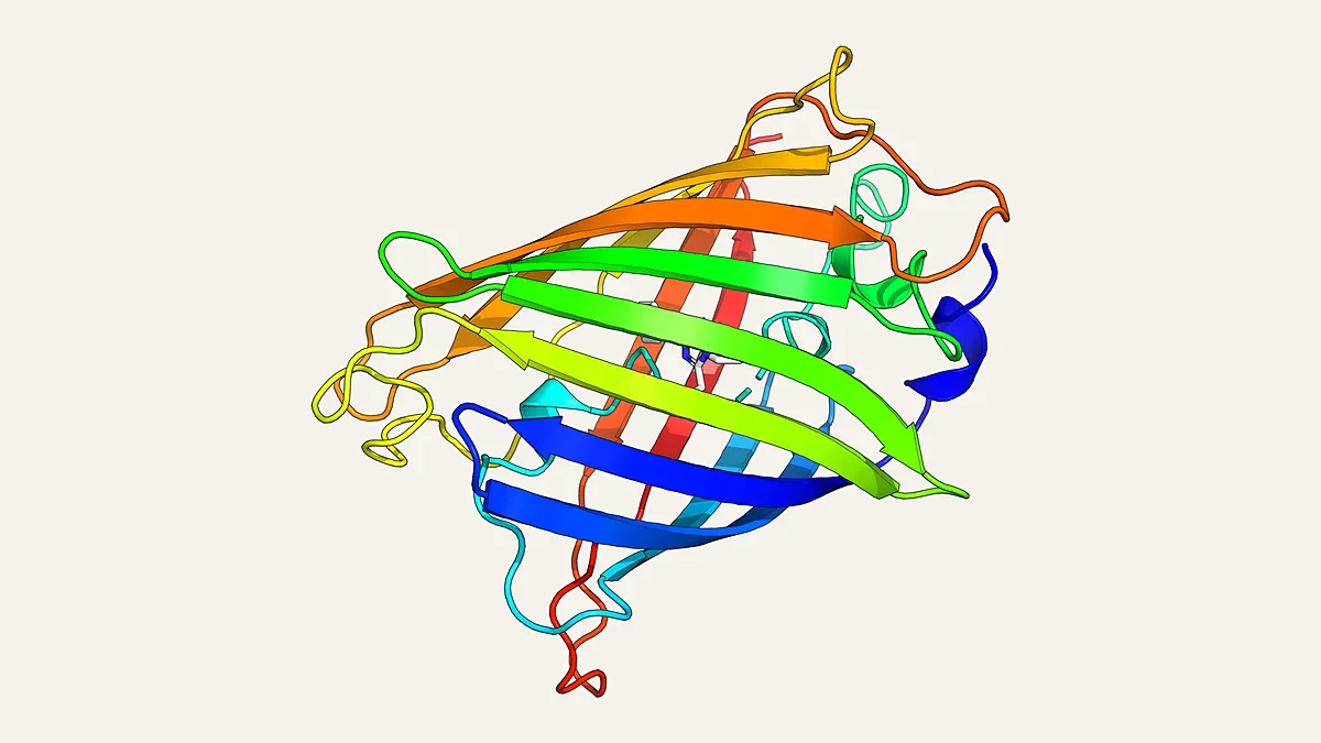
Physics-based pocket design gives de novo proteins pre-organised binding sites for chromophores
Researchers at the University of Bristol, with collaborators at the NNF Center for Protein Design at the University of Copenhagen, the University of Sheffield and the University of Manchester, reported in Nature Communications, published online on 19 October, a rapid physics-based computational method for building small-molecule binding pockets into de novo designed proteins.
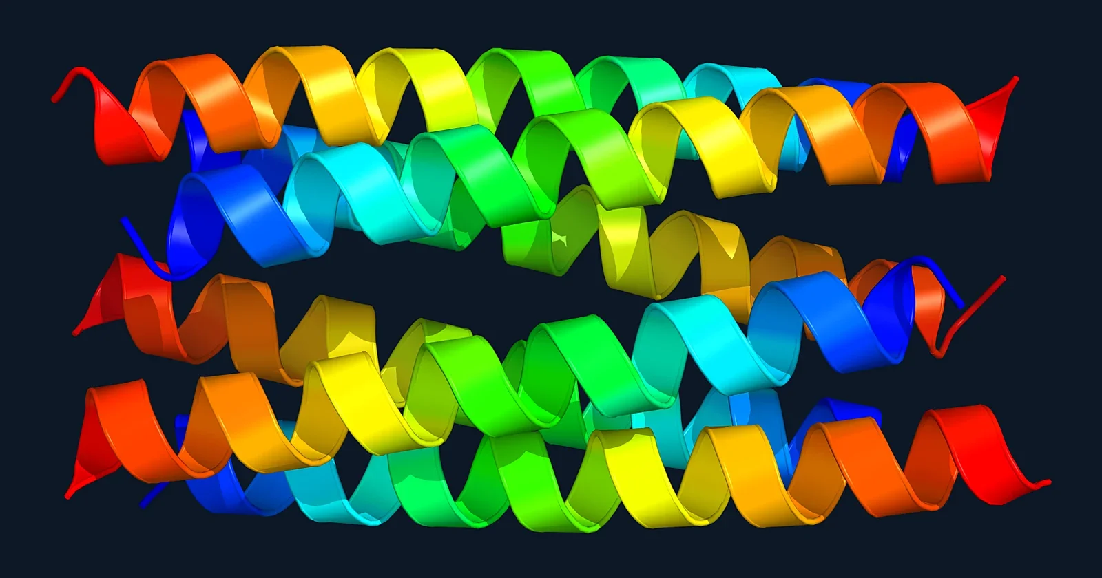
Summary. Researchers at the University of Bristol, with collaborators at the NNF Center for Protein Design at the University of Copenhagen, the University of Sheffield and the University of Manchester, reported in Nature Communications, published online on 19 October, a rapid physics-based computational method for building small-molecule binding pockets into de novo designed proteins. Deep-learning methods can now generate new protein structures quickly, but designing proteins that bind a chosen small molecule tightly, specifically and predictably remains difficult. The new method generates binding pockets that are isosteric with the target molecule, meaning matched to its shape and size, and chemically complementary to it.
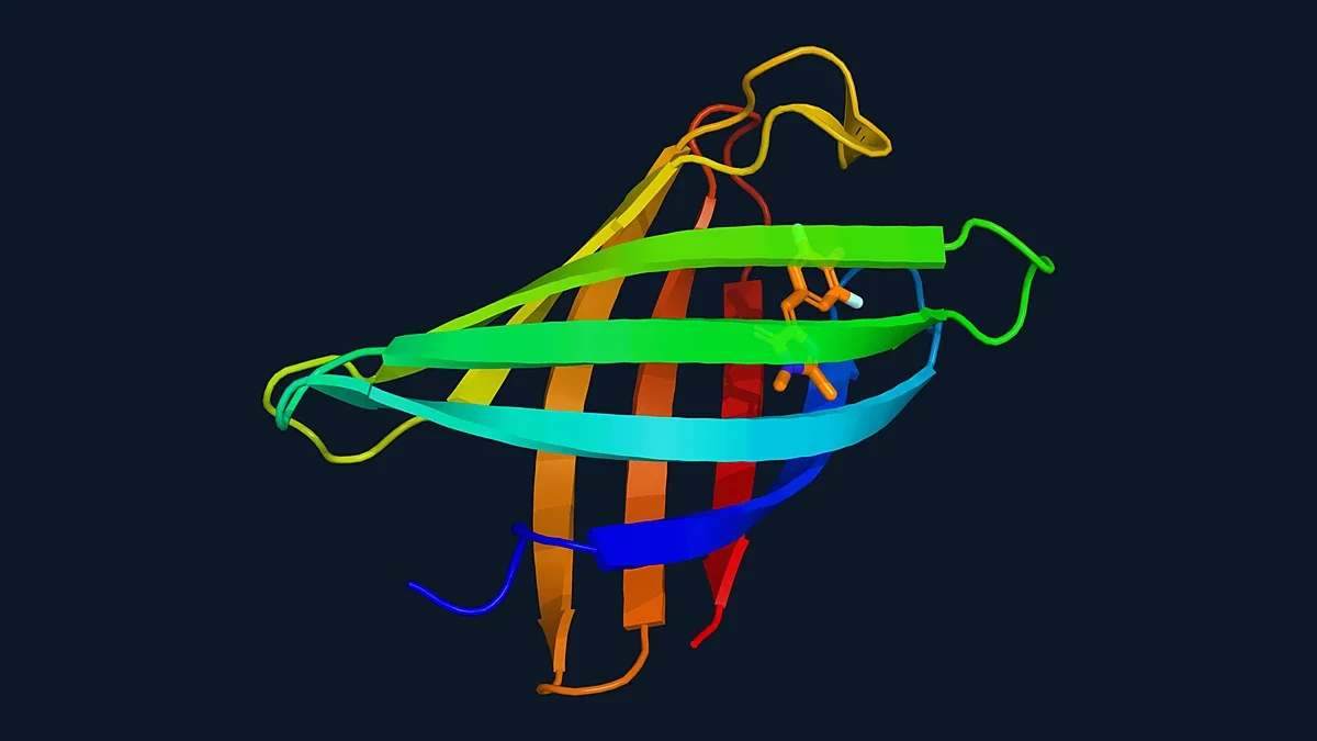
Research context (RUO). The team tested the pipeline experimentally by building and characterizing binding proteins for several synthetic and natural chromophores, coloured molecules that absorb visible light. Only single-digit numbers of designs had to be evaluated, and the pipeline delivered stable proteins whose binding sites were already pre-organised in the ligand-free (apo) state, as confirmed by X-ray crystallography, and which bound their targets with micromolar affinities or better. To show the scope of the approach, the authors built two selective chromophore-binding sites into a single two-domain de novo protein and coupled them so that energy could be transferred between the two sites in a controlled way. They also developed a small de novo binding protein that could be used in live mammalian cells to visualise sub-cellular structures. X-ray data were collected at the Diamond Light Source and the European Synchrotron Radiation Facility, and the paper is open access. The work is basic protein-design research with no clinical component. For protein-engineering and chemical-biology laboratories, it suggests that small-molecule binders can be obtained from very few designs when pocket shape and chemistry are matched to the ligand from the start.