Benchmark finds AI cofolding models can place a ligand correctly while mispredicting the protein's conformation
Researchers at Freie Universität Berlin reported in the Journal of Chemical Information and Modeling, published online on 20 October, a benchmark of four AI cofolding models that predict protein-ligand complexes: AlphaFold3, RoseTTAFold3, Boltz-2 and Chai-1.
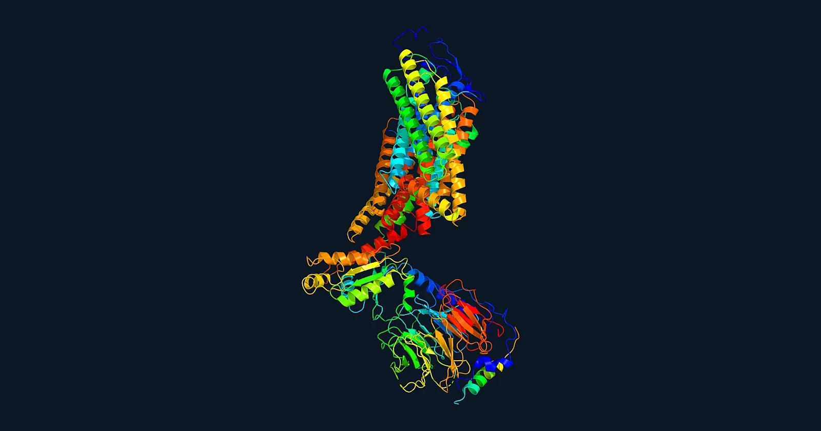
Summary. Researchers at Freie Universität Berlin reported in the Journal of Chemical Information and Modeling, published online on 20 October, a benchmark of four AI cofolding models that predict protein-ligand complexes: AlphaFold3, RoseTTAFold3, Boltz-2 and Chai-1. The study asked whether a correctly placed ligand also means the surrounding protein was predicted in the right conformation. The authors found that the two outcomes are systematically decoupled: a ligand can sit close to its experimental position while the protein around it adopts the wrong functional state.
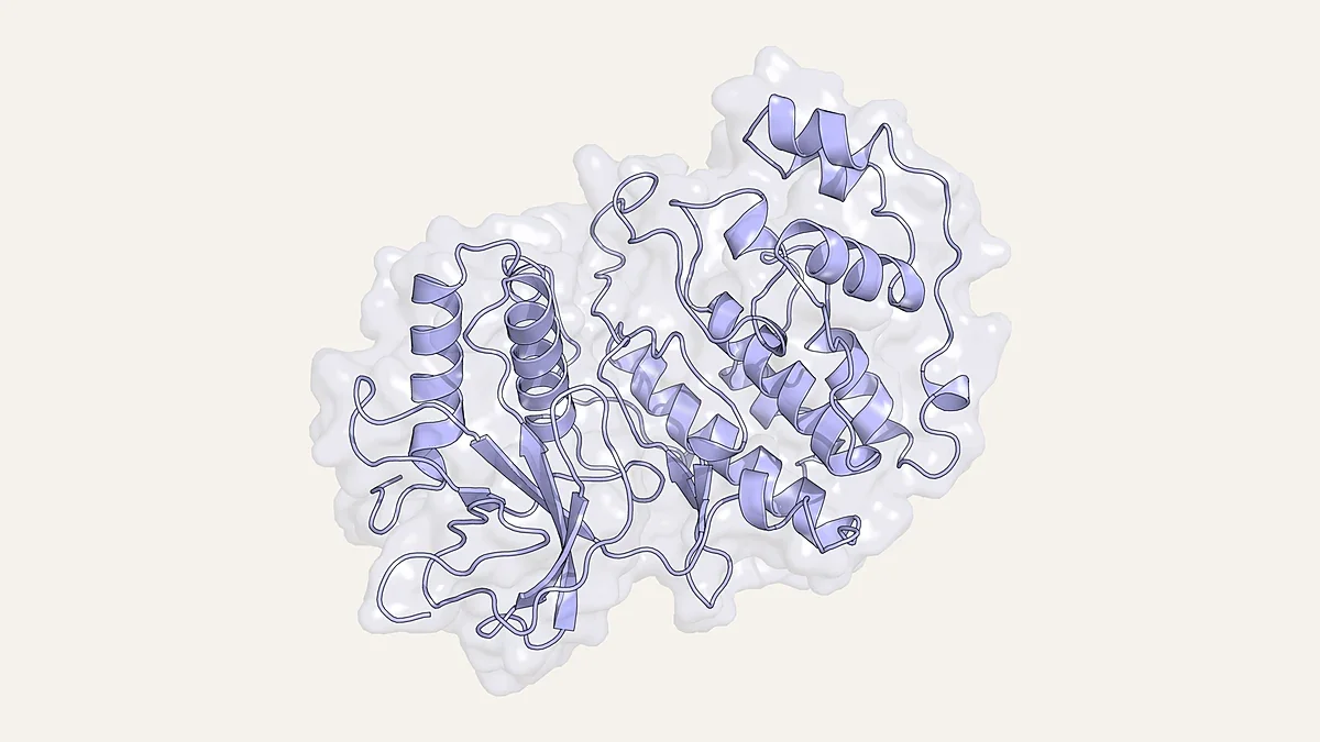
Research context (RUO). The team used kinases and Class A G-protein-coupled receptors (GPCRs) as test systems, because both families depend on well-defined conformational states. To make the test prospective, they evaluated complexes released after the training cutoffs of all four models, together with one earlier nucleotide-bound structure as a control. State-defining features, such as the kinase activation loop and DFG/DYG geometry, and intracellular GPCR regions such as the third intracellular loop (ICL3), were frequently mispredicted even when the ligand root-mean-square deviation (RMSD) was low. The models remodelled the binding pocket locally around the ligand, but that adaptation rarely carried through to the correct global conformation. Adding state-annotated templates and filtered multiple sequence alignments improved conformational recovery in some cases and had little effect in others. Model confidence scores tracked ligand placement but could not report whether the local pocket geometry was right, and the single allosteric complex in the set was misplaced by all four models, even with explicit guidance. The paper is a computational benchmarking study with no experimental or clinical component. For structural-biology and modelling laboratories, it is a reminder to check the predicted protein state separately from ligand placement when using cofolding models.