Time-resolved AFM shows how a second peptide crystal layer nucleates on top of the first
An open-access Advanced Functional Materials study published October 22, 2026 used time-resolved atomic force microscopy to follow how a second layer of peptide crystals nucleates and grows on a first peptide layer on graphite.
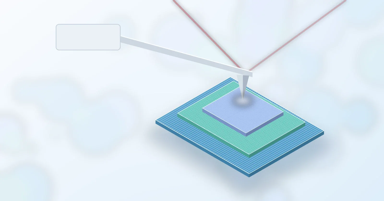
Short peptides that stick to flat solid surfaces can organize themselves into ordered, crystal-like films. A new study now shows, step by step, how a second crystalline layer begins to form on top of the first. The paper, "Beyond Primary Nucleation: Molecular Basis of Peptide Crystal Secondary Nucleation at Soft Interface," was published open access in Advanced Functional Materials on October 22, 2026, by Linhao Sun, Abilesh Kumar and Ayhan Yurtsever of the WPI Nano Life Science Institute at Kanazawa University. [1]
The peptide and the surface
The team worked with GrBP5, a graphite-binding dodecapeptide. A 2012 ACS Nano paper showed that dodecapeptides of this kind, selected by phage display, self-assemble on graphite into long-range-ordered nanostructures, and that simple mutations can tune their binding, surface aggregation and growth kinetics. [2] In 2023, part of the same Kanazawa group used in situ atomic force microscopy (AFM) in liquid to follow GrBP5 forming its first two layers on graphite. They found that the first layer forms homogeneously, while the second is mostly heterogeneous, triggered by crystalline defects in the layer below. [3]
Watching nucleation happen
The new work focuses on secondary nucleation: new crystals forming on an existing assembled surface rather than directly from solution. A 2018 review describes it as new peptide aggregates nucleating on the surfaces of existing fibrils of the same peptide, a pathway that has emerged over the past two decades as a major route for aggregate growth in solution. [4] The authors note that those studies largely relied on population-averaged measurements. [1]
Using time-resolved 3D-AFM and high-speed AFM in liquid, plus infrared spectroscopy, the team reports: [1]
- A critical concentration of about 5 micromolar for a second layer to appear. Below that, the first layer dominates. At 250 micromolar a third layer forms.
- Nuclei smaller than 5 nm first appear at grain boundaries in the first layer and at step edges of the graphite. Mobile "nanostrings" act as a temporary reservoir of peptide.
- A layer spacing of about 0.6 nm. The second layer has a wider lateral repeat (4.9 nm versus 3.2 nm), so three of its rows line up with five of the first layer's.
- Unstable growth directions shrink and disappear over several hours, leaving nanowires at stable 60° offsets.
Charge changes the speed
The group also compared GrBP5 with two mutants, one carrying two extra negatively charged residues and one carrying two extra positively charged residues. At 50 micromolar, the second layer of the positively charged mutant grew fastest. The authors attribute this to faster anchoring on the first peptide layer. [1] They also showed cross-seeding: one sequence's crystals can grow on a first layer made of another, giving hybrid layered crystals. [1] A 2025 Journal of Colloid and Interface Science paper from overlapping authors had already reported stacked 3D peptide crystals built layer by layer on graphite, MoS2 and mica. [5]
Why it matters for peptide research
The authors frame the results as design rules for peptide crystal engineering and layered bio/nano interfaces, with possible uses in biosensing and bioelectronics. As next steps, they plan to test whether the early nanoclusters resemble droplets formed by liquid–liquid phase separation. [1] For labs working on peptide self-assembly, the study gives a rare molecular-scale, time-resolved view of how each new layer starts.
Related: Self-assembling amyloid-forming peptide design · Phage-display peptides that block ice nucleation · What are peptides? · Basic terms · Peptide Academy
All BioLabs Research products are sold strictly for laboratory research use only. Not for human or veterinary use. This article summarizes published research and makes no claims about any BioLabs product.
- Sun L., Kumar A., Yurtsever A., "Beyond Primary Nucleation: Molecular Basis of Peptide Crystal Secondary Nucleation at Soft Interface," Advanced Functional Materials, published October 22, 2026 (open
- So C.R., Hayamizu Y., Yazici H., Gresswell C., Khatayevich D., Tamerler C., Sarikaya M., "Controlling Self-Assembly of Engineered Peptides on Graphite by Rational Mutation," ACS Nano, 2012
- Yurtsever A., Sun L., Hirata K., Fukuma T., Rath S., Zareie H., Watanabe S., Sarikaya M., "Molecular Scale Structure and Kinetics of Layer-by-Layer Peptide Self-Organization at Atomically Flat Solid S
- Törnquist M., Michaels T.C.T., Sanagavarapu K., Yang X., Meisl G., Cohen S.I.A., et al., "Secondary nucleation in amyloid formation," Chemical Communications, 2018
- Hu J., Yurtsever A., Wang D., Sun L., "Formation of stacked 3D crystals of self-assembled peptides via mimicking construction of layered natural minerals," Journal of Colloid and Interface Science, 20